 Mistake Master
Plasma membranes
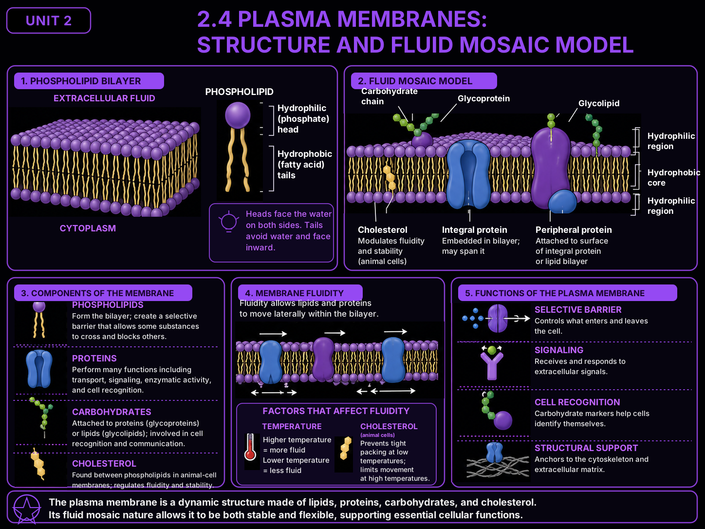
Every cell is wrapped in a plasma membrane, and the model that describes it is the fluid mosaic. "Mosaic" because the membrane is a patchwork of many parts — a phospholipid bilayer studded with proteins, cholesterol, and surface carbohydrates. "Fluid" because those parts are not locked in place: the lipids and many proteins drift sideways, so the whole sheet behaves like a two-dimensional liquid rather than a rigid wall. Read the structure — amphipathic phospholipids with water-loving heads facing out and water-fearing tails tucked inside — and the membrane's job as a selective barrier follows directly.
§1
The big idea: a fluid mosaic, not a solid wall.
▸
The plasma membrane is described by the fluid mosaic model, and both words matter. Mosaic means the membrane is a patchwork of different parts — a phospholipid bilayer as the base sheet, with proteins, cholesterol, and carbohydrate chains scattered through and across it. Fluid means those parts are not nailed down: individual phospholipids and many of the proteins drift sideways within the layer, so the membrane behaves like a thin two-dimensional liquid.
This is the single most misremembered point in the topic. A membrane is not a rigid, static, solid wall with everything locked in a fixed grid. It is dynamic and constantly in motion — molecules slide past one another, proteins wander, and the whole sheet flexes and self-heals. Picture a crowd of people milling on a plaza, not bricks cemented in a wall.
The base of the mosaic is the bilayer — two sheets of phospholipids arranged tail-to-tail. Because a phospholipid is amphipathic (one end loves water, the other flees it), it can only sit one way in a watery cell: hydrophilic heads facing the water inside and outside the cell, hydrophobic tails buried in the middle. Get that orientation right and everything else about the membrane — how it forms, why it is selective, why it stays fluid — starts to make sense.
§2
How the bilayer builds itself: phospholipid orientation.
▸
A single phospholipid has two chemically opposite ends, and that split personality is what forces the bilayer into shape. Walk through the logic step by step.
- The head loves water. The phosphate head group is polar and charged — it is hydrophilic, so it is attracted to water and settles wherever there is watery fluid.
- The tails flee water. The two fatty-acid tails are nonpolar — they are hydrophobic, so they are pushed away from water and try to hide from it.
- A cell has water on both sides. There is watery cytoplasm inside the cell and watery fluid outside. So there is water above and below wherever the membrane sits.
- Two layers form, tail-to-tail. The only stable arrangement is a double layer: heads of one sheet face the outside water, heads of the other sheet face the inside water, and both sets of tails point inward toward each other — shielded from water in the oily core. That is the bilayer.
- It self-assembles. Nobody has to arrange the phospholipids. Drop them in water and they snap into heads-out, tails-in on their own, because that is the lowest-energy way to satisfy both ends at once.
Hold onto the correct picture: heads out toward the water on both surfaces, tails buried in the middle. Flipping that — tails facing the water, or heads pointing inward — is one of the most common and costly errors on this topic.
§3
The parts of the mosaic.
▸
Quick reference card for the components embedded in and around the bilayer. For each, read the structure and the function it fits — and notice that everything is set within a layer that stays fluid.
§4
Why fluid matters, and how structure fits function.
▸
The mosaic being fluid is not a trivia point — the membrane could not do its jobs if it were a frozen solid. Here is what the fluidity buys, and how each structural feature is matched to a function.
Lipids and proteins drift. Within the bilayer, individual phospholipids swap places millions of times a second, and many embedded proteins wander laterally across the cell surface. This constant motion is the normal, healthy state — not a sign of damage. Because the sheet is fluid, it can bend around a dividing cell, pinch off vesicles, and reseal when punctured.
Cholesterol is the fluidity buffer. In animal membranes, cholesterol molecules sit between the phospholipids and act as a thermostat. At high temperature they restrain the phospholipids and keep the membrane from getting too loose; at low temperature they space the phospholipids apart and keep the membrane from packing into a stiff gel. The structure — a rigid ring wedged among flexible tails — fits the function of holding fluidity in a workable range.
The bilayer is a selective barrier. The hydrophobic core repels water-soluble ions and molecules, so they cannot slip straight through. That is the structure that fits the membrane's core function: keeping the inside and outside chemically different. Small nonpolar molecules can dissolve through, but most everything else needs help.
Embedded proteins are the doors. Because the plain bilayer blocks charged and large molecules, the membrane studs itself with transport proteins — channels and carriers that span the layer and provide controlled passageways. The structure (a protein tunnel through the oily core) fits the function (selective, regulated transport). Other membrane proteins act as receptors, enzymes, and anchors.
Surface carbohydrates are ID tags. Short sugar chains attached to outer proteins and lipids project from the cell's outside face. Their structure — distinctive branching patterns facing outward — fits a recognition function: they let cells identify one another and distinguish self from foreign.
Read structure to function. Every feature earns its place. Amphipathic phospholipids → a self-sealing barrier; a hydrophobic core → selectivity; embedded proteins → controlled transport; cholesterol → stable fluidity; surface sugars → recognition. Name the structural feature and you can name the job — that is the heart of Topic 2.4.
§5
3 mistakes that cost real points.
▸
“The membrane is a static, solid wall with everything locked in place.”
This is the signature misconception of the topic. Students picture the membrane as a rigid sheet of bricks — molecules cemented in a fixed grid, nothing moving. But the model is called fluid mosaic for a reason: phospholipids and many proteins drift sideways constantly, and that motion is the normal, healthy state. A solid membrane could not bend, fuse vesicles, or reseal after a puncture — a cell needs it to be a living, moving liquid, not a wall.
Fix. Picture a crowd milling on a plaza, not bricks in a wall. If a question implies the membrane is rigid, static, or frozen in place, treat that as a red flag — membranes are dynamic by design.
“The phospholipid tails face the water and the heads point inward.”
This orientation error flips the whole bilayer backward. The rule is fixed by chemistry: heads are hydrophilic and go toward water; tails are hydrophobic and hide from it. Since a cell has water on both sides, the only stable layout is heads facing out toward the fluid on each surface and tails buried in the middle, away from water. Putting tails in the water — or drawing a single layer instead of two — contradicts the chemistry and is a classic lost point.
Fix. Say it every time: heads out to the water, tails tucked in. Two layers, tail-to-tail. If your picture has tails touching water, you've drawn it upside down.
“The membrane's parts are just there; their structure has nothing to do with what the membrane does.”
This is the structure–function disconnect applied to the membrane. Students treat “the bilayer has a hydrophobic core” and “the membrane is selectively permeable” as two unrelated facts. But the oily core is the reason water-soluble molecules are blocked; the embedded proteins are the reason chosen molecules can still cross; the surface sugars are the reason cells recognize each other. Structure and function are one idea, not two.
Fix. For every membrane component, finish the sentence “this feature does this job because…” If you can't, you've memorized a label without understanding the structure behind it.
§6
Skill Check.
▸
Ten scenarios. Pick the chips that match your answer, then check. A scenario marks complete the first time every part is right. Progress saves on this device.